Retinal Detachment
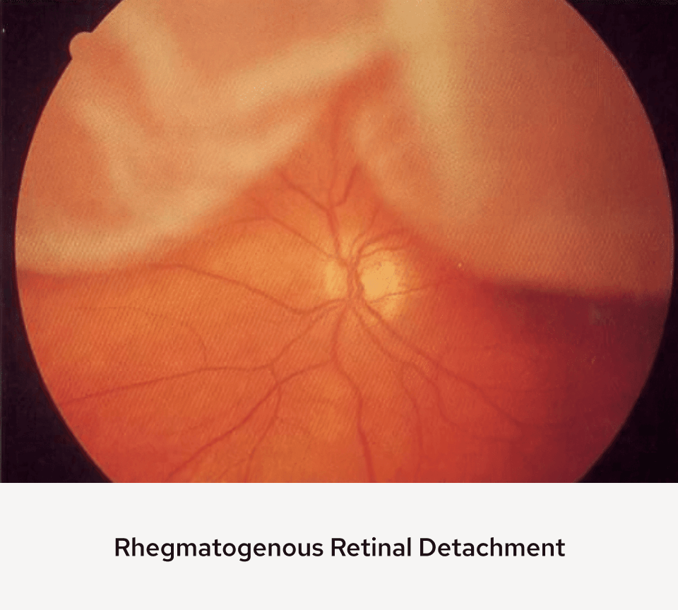
The retina is the thin film that covers the back of the eye. It’s able to collect light and create electrical signals that are then sent to the brain and allow us to see. A retinal detachment occurs when a tear or hole in the retina allows the liquid vitreous to get under the retina and accumulate. The retina separates from the back wall of the eye. When the retina is detached, it does not work and people notice a gradually enlarging veil or dark shadow in the peripheral visual field. When the detachment extends into the central part of the retina (macula) the central vision is lost.
Retinal detachments may be fixed in the office in some selected cases by using a gas bubble and laser treatment called pneumatic retinopexy. More complex detachments commonly require scleral buckling or vitrectomy surgery in a surgery center with anesthesia. In scleral buckling surgery, the retinal tear or tears (there may be several or many) are treated with cryotherapy (freezing treatment) and a band is sutured around the eye that indents the eye wall inward against the detached retina. This bump seals the retinal tears and the fluid under the retina is absorbed by the body. Thus, the retina becomes reattached to the back wall of the eye.
Another way to fix retinal detachments is to perform vitrectomy surgery. The vitreous is removed using small needle-like microinstruments. The tear is identified and treated with laser and the fluid under the retina is removed with a straw-like instrument. All of the fluid in the eye is then removed, and a gas bubble is placed in the eye. The bubble acts as an internal splint keeping the retina in place while the laser heals. The body absorbs the gas bubble slowly over a couple of weeks and the eye replaces the bubble with its own fluid. The vitreous does not grow back and is not necessary for the normal functioning of the eye. Your retina surgeon will decide which of these procedures is the best way to fix each retinal detachment.
Schedule a Consultation for Retinal Detachment
The retina specialists of AVRUC provide expert care for Retinal Detachment. With convenient retina center locations in Carmel, Bloomington, Muncie, and Avon, we welcome patients from the greater Indianapolis area, Evansville, Lafayette, Terre Haute, and beyond.